
Brain Imaging of Blast Injury
By:
Dr. Carol Henricks
On
13/02/2025Summary:
When a physician chooses a test to evaluate an injury or illness, it is a specific test that will demonstrate the pathology that is of concern. An ER physician who is concerned that a patient has had a stroke would NOT do an x-ray of the skull.
Head CT Scan
An ER physician who is evaluating a patient with concussion is likely to do a head CT scan to evaluate for skull fracture or intracranial bleed, but if that test is “unremarkable” it DOES NOT rule – out concussion.
Head CT Scan
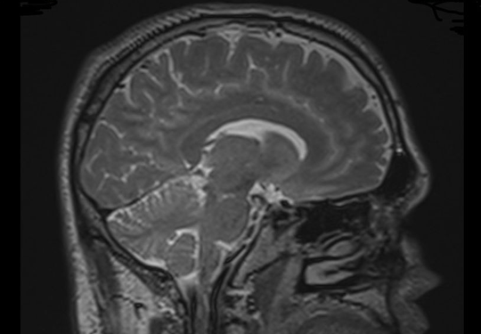
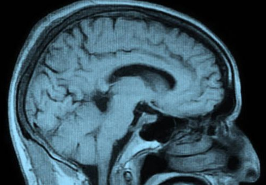
Normal Brain MRI
A routine MRI of the brain, even a contrast study, does not adequately evaluate for concussion or blast brain injury. Many times, a brain MRI that is done without special applications is interpreted as normal or normal for age but there is actually significant brain injury.
"Normal MRI – DTI”
A concussion or blast injury causes a diffuse injury throughout the brain: multiple small lesions that are not detectable at a gross anatomical level. The neuropathology of the injury must be evaluated in a different way. The genius of brain MRI with DTI application is that physiological changes in the brain which represent injury are demonstrated in conjunction with tractography (the drawing of fiber tracts in the brain). Interruptions in the fiber tracts are consistent with injury at that location. Damaged fiber tracts interfere with network functions of the brain.
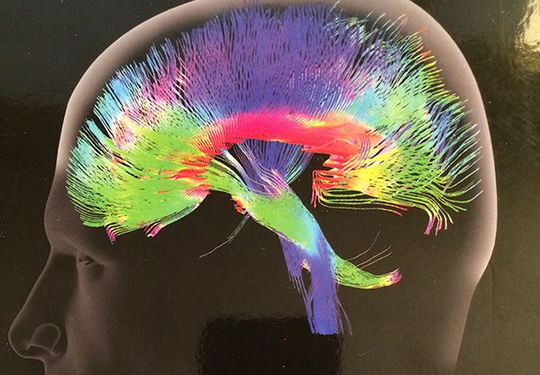
Normal MRI-DTI

NORMAL MRI-DTI
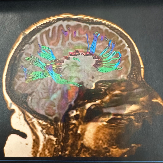
ABNORMAL MRI-DTI
“Blast Injury Demonstrated with MRI -DTI"
We cannot continue to misdiagnose veterans who have suffered concussion and blast injury because they are not having the most specific, diagnostic testing done. You cannot heal if you have never been properly diagnosed.